


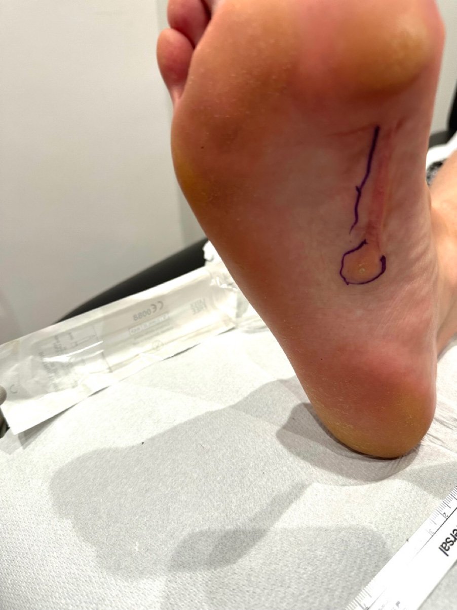
As a Podiatrist with a clinical interest in connective tissue disorders, I recently had the opportunity to treat a 30-year-old woman suffering from Ledderhose disease. The following blog/case study involved the use of focused Extracorporeal Shock Wave Therapy (fESWT) to address recurrent painful plantar fibromas in the plantar fascia of her right foot. In prior treatments, surgical excision and radiation therapy had provided temporary relief, but the fibroma eventually regrew to its original size. This blog aims to shed light on the patient’s experience and the remarkable outcomes observed following focused shockwave as part of her treatment plan. (Focused shockwave is different to and should not be confused with another form of shockwave called radial. Radial shockwave is ineffective for plantar fibroma treatment).
Understanding Ledderhose Disease:
Ledderhose disease, also known as plantar fibromatosis, is a rare connective tissue disorder. It is characterised by the development of benign fibrous growths (plantar fibromas) in the plantar fascia, the thick tissue running along the bottom of the foot. These fibromas can cause pain, and discomfort, and limit mobility, significantly impacting a person’s quality of life.
Past medical history.
Before the patient sought treatment with focused shockwave, she had experienced surgical excision and radiation therapy. While the latter treatment options provided temporary relief, the fibroma and discomfort ultimately returned, leading the patient to explore alternative treatment options. It was during this search that she discovered focused shockwave.
Focused Shockwave: A Non-Invasive Solution:
Focused Extracorporeal Shock Wave Therapy is a relatively new treatment modality that utilises high-energy shock waves to target affected tissues. In this case, the patient’s plantar fibroma was treated using focused shockwave therapy. Prior to commencing treatment, an ultrasound scan was performed to eliminate the possibility of malignancy and to measure the dimensions of the fibroma accurately, and map out the positions of nearby nerves. The baseline measurements of the fibroma indicated a height from top to bottom of 6.1 mm and a length of 9.3 mm.
Patient Experience and Treatment Outcomes:
The treatment plan consisted of two 30-minute appointments, that involved ultrasound evaluation, followed by 15 minutes of focused shockwave treatment. During the treatment the patient experienced transient discomfort graded as 4/10 on the visual analogue pain scale, on a couple of occasions during treatment at certain angulations of the hand-piece the discomfort rose to a 7/10 for 1-2 seconds. Immediately after treatment, the patient’s discomfort level reduced to the pre-treatment baseline of 6/10. The day following treatment the patient experienced several brief occurrences of increase in discomfort, graded as 7 out of 10 on the Visual Analog Scale. However, by day 5, the discomfort had substantially reduced to 2 out of 10 on the VAS, and by day 6, it completely subsided, with the patient now reporting a 0 out of 10 pain score. The patient did not report any side effects associated with the therapy. During the second treatment session performed 6 days after the first, the patient experienced transient discomfort graded as 4 out of 10 on the visual analogue scale, immediately after treatment the patient scored 0 out of 10 on the visual analogue scale.
Remarkable Changes: Measurement and Patient Satisfaction:
The patient’s delight was evident when she discovered that focused shockwave was a viable option after radiation therapy. Unlike her previous radiation treatment, she did not experience any dryness or redness in the treated area. The positive outcomes of focused shockwave were also visible through ultrasound evaluation, which revealed a significant reduction in fibroma dimensions. The top-to-bottom height had decreased from 6.1 mm to 3.2 mm, and the length reduced from 9.3 to 5 mm after the first treatment.
This case study demonstrates the potential efficacy of focused Extracorporeal Shock Wave Therapy in managing plantar fibromatosis in Ledderhose disease. The patient’s experience and treatment outcomes highlight the non-invasive nature of focused shockwave, its ability to alleviate pain and discomfort, and the absence of adverse side effects.
How might focused shockwave treatment work for fibromas at a cellular level?
I am afraid the next section of this blog is going to be a little bit science-heavy and not for everyone, if understanding the finer detail isn’t for you feel free to skip to the conclusion.
Therapeutic Effects of focused shockwave on fibromas.
Focused shockwave involves the delivery of high-energy shockwaves to the affected area, promoting healing and reducing fibrous tissue formation. The therapeutic effects of focused shockwave on fibroma tissue are multi-faceted and encompass several key aspects:
1. Promotion of Collagen Remodelling:
Focused shockwave stimulates the up-regulation of collagenase enzymes, leading to increased degradation of abnormal collagen fibres found in the lumps. This process facilitates the remodelling of fibroma tissue, reducing its size, improving its appearance and improving the overall texture of the skin.
2. Enhancement of Angiogenesis:
Focused shockwave has been shown to promote the growth of new blood vessels around the fibroma tissue, a process called angiogenesis. This angiogenic effect is beneficial as it increases blood supply to the area, facilitating the transport of vital nutrients and oxygen to support the healing process. Improved vascularisation also aids in the removal of waste products, further reducing inflammation and fibrosis.
3. Reduction of Fibrosis:
Focused shockwave has anti-fibrotic effects, decreasing the excessive proliferation of fibroblasts and myofibroblasts cells, which are the key cellular players in fibroma formation. By inhibiting fibrosis, focused shockwave helps to prevent the excessive production and deposition of collagen, resulting in a decrease in fibroma size and discomfort.
4. Mechanical Disruption:
The rapid application of high-energy shockwaves delivered during focused shockwave causes mechanical disruption of the fibroma tissue. This fragmentation leads to the breakdown of the abnormal collagen fibres and stimulates the activation of cellular signalling pathways involved in tissue regeneration and remodelling.
5. Cellular Signalling Modulation:
Focused shockwave is believed to modulate the expression of various signalling molecules, such as transforming growth factor-beta (TGF-β) and vascular endothelial growth factor (VEGF). By altering the expression of these molecules, ESWT can attenuate inflammatory responses, promote collagen degradation, and drive angiogenesis, ultimately facilitating fibroma reduction or resolution.
6. Stimulation of Cellular Metabolic Activity:
Focused shockwave triggers an increase in cellular metabolic activity by promoting ATP production within the target tissue. This augmented cellular activity enhances reparative processes by accelerating cell division, migration, and proliferation at the site of the fibroma.
Conclusion:
Focused shockwave offers a novel therapeutic approach for the management of plantar fibroma, with its desirable effects on collagen remodelling, angiogenesis, and fibrosis reduction. While the exact mechanisms of action remain to be fully understood, mechanical disruption, cellular signalling modulation, and stimulation of cellular metabolic activity are proposed as key contributors. These findings highlight the potential of focused shockwave as an effective and non-invasive treatment modality, providing hope for individuals seeking relief from plantar fibroma. Furthermore, focused shockwave has an excellent safety profile making it the treatment of choice for those that have had previous radiation treatment or surgery. Unlike radiation therapy, there is no limit to how many times a fibroma can be treated.
What is the long-term outlook for our case study?
It is highly likely the size of the fibroma will reduce further from her second treatment. A previously published study entitled “ High energy focused extracorporeal shockwave therapy reduces pain in plantar fibromatosis (Ledderhose disease.)” showed that patient’s discomfort at 3 months post-treatment reduced to a 1 or 0 on the VAS following focused shockwave. Broadly speaking focused shockwave appears equally as effective as radiation therapy. Radiation and focused shockwave can sometimes be curative of individual fibroma or can give substantial improvement lasting years.
References:
-
Falanga V, et al. Mechanisms of action of shockwave therapy in soft tissue conditions. Eur Cell Mater. 2018 Apr 6;35:59-84.
-
Liu N, et al. Extracorporeal shockwave therapy: A potential novel therapeutic option for keloids. J Cosmet Dermat3. Knobloch K, Vogt PM. High-energy focussed extracorporeal shockwave therapy reduces pain in plantar fibromatosis (Ledderhose’s disease). BMC Res Notes. 2012 Oct 2;5:542. doi: 10.1186/1756-0500-5-542. PMID: 23031080; PMCID: PMC3637402. View link here